
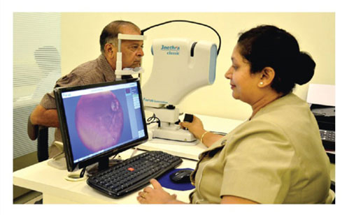
A photo of the patient’s Retina is taken by an Ophthalmic Technician, Nurse or Medical assistant using an easy-to-use, non-Mydriatic Fundus camera. This process is quick , non invasive and does not require any dilatation.
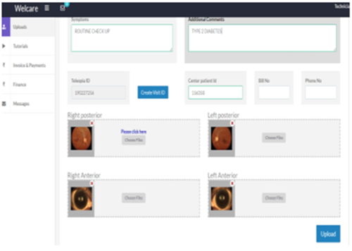
Once the Retinal photos are taken, images are uploaded to Welcare Cloud Based Software and forwarded to Teleophthalmology platform along with patient’s medical history information.
The transfer is secure and encrypted.
Images are permanently archived and accessible Support for all image formats including DICOM
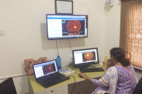
Images are interpreted remotely by our Ophthalmologists and Retina Specialists using established international classification guidelines.
Image enhancement and annotation done.
Built-in interpretation modules for diabetic retinopathy, glaucoma, AMD.
Once the ophthalmologist interpretation is complete, the patient’s diagnostic report is generated indicating any abnormalities with recommendations for follow-up or treatment within 30 minutes.
Easy-to-understand report format.
Report is transmitted back to the Technician and their provider can be accessed online.